Accurate and efficient gesture spotting via pruning and subgesture reasoning

Accurate and E?cient Gesture Spotting via Pruning and Subgesture Reasoning
Jonathan Alon,Vassilis Athitsos,and Stan Sclaro?
Computer Science Department
Boston University
Boston,MA02215,USA
Abstract.Gesture spotting is the challenging task of locating the start
and end frames of the video stream that correspond to a gesture of inter-
est,while at the same time rejecting non-gesture motion patterns.This
paper proposes a new gesture spotting and recognition algorithm that is
based on the continuous dynamic programming(CDP)algorithm,and
runs in real-time.To make gesture spotting e?cient a pruning method
is proposed that allows the system to evaluate a relatively small num-
ber of hypotheses compared to CDP.Pruning is implemented by a set
of model-dependent classi?ers,that are learned from training examples.
To make gesture spotting more accurate a subgesture reasoning process
is proposed that models the fact that some gesture models can falsely
match parts of other longer gestures.In our experiments,the proposed
method with pruning and subgesture modeling is an order of magnitude
faster and18%more accurate compared to the original CDP algorithm.
1Introduction
Many vision-based gesture recognition systems assume that the input gestures are isolated or segmented,that is,the gestures start and end in some rest state. This assumption makes the recognition task easier,but at the same time it limits the naturalness of the interaction between the user and the system,and therefore negatively a?ects the user’s experience.In more natural settings the gestures of interest are embedded in a continuous stream of motion,and their occurrence has to be detected as part of recognition.This is precisely the goal of gesture spotting:to locate the start point and end point of a gesture pattern,and to classify the gesture as belonging to one of predetermined gesture https://www.wendangku.net/doc/0f17628056.html,-mon applications of gesture spotting include command spotting for controlling robots[1],televisions[2],computer applications[3],and video games[4,5].
Arguably,the most principled methods for spotting dynamic gestures are based on dynamic programming(DP)[3,6,7].Finding the optimal matching between a gesture model and an input sequence using brute-force search would involve evaluating an exponential number of possible alignments.The key ad-vantage of DP is that it can?nd the best alignment in polynomial time.This is This research was supported in part through U.S.grants ONR N00014-03-1-0108, NSF IIS-0308213and NSF EIA-0202067.
677 (5S)
750 (5E)807 (6S)873 (6E)
10
20
30
40
50
677 (5S)
750 (5E)807 (6S)873 (6E)
10
20
30
40
50
(a)(b)(c)
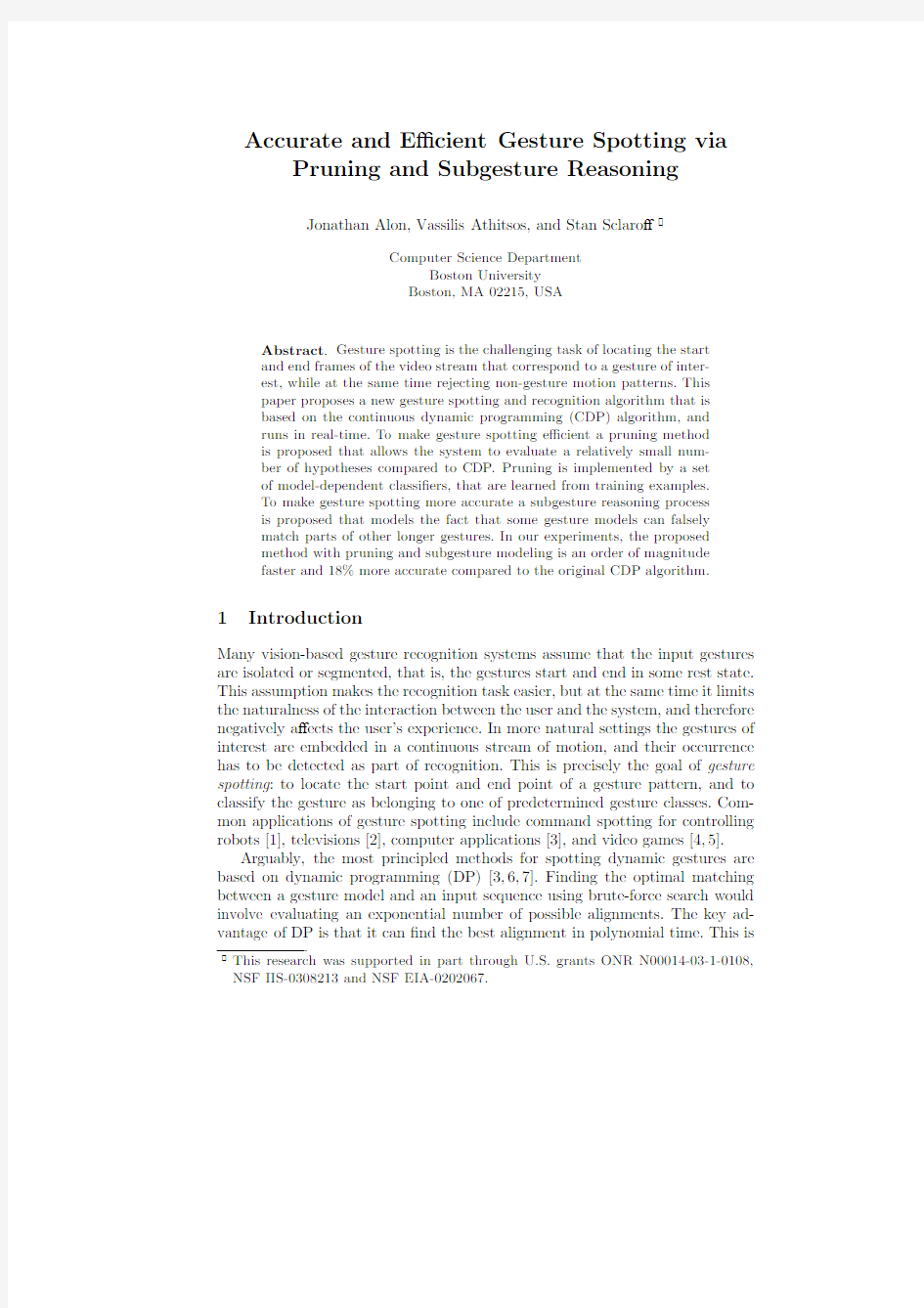
Fig.1.Pruning(a,b):example dynamic programming table for matching input stream (x axis)to a model gesture for the digit“6”(y axis).Likely observations are represented by black cells in the table(a).The cells remaining after pruning(b).In this example 87%of the cells(shown in white)were pruned.Subgesture reasoning(c):example false detection of the digit“5”,which is similar to a subgesture of the digit“8”. achieved by reducing the problem of?nding the best alignment to many subprob-lems that involve matching a part of the model to parts of the video sequence. The main novelty of our method is a pruning technique that eliminates the need to solve many of these subproblems.As a result,gesture spotting and recog-nition become both faster and more accurate:faster because a smaller number of hypotheses need to be evaluated;more accurate because many of the hy-potheses that could have led to false matches are eliminated at an early stage. In Figure1(b)the number of hypotheses evaluated by the proposed algorithm is proportional to the number of black pixels,and the number of hypotheses that are evaluated by a standard DP algorithm but are pruned by the proposed algorithm is proportional to the number of white pixels.
A second contribution of this paper is a novel reasoning process for decid-ing among multiple candidate models that match well with the current portion of the input https://www.wendangku.net/doc/0f17628056.html,paring the matching scores and using class speci?c thresholds,as is typically done[3,6],is often insu?cient for picking out the right model.We propose identifying,for each gesture class,the set of“subgesture”classes,i.e.,the set of gesture models that are similar to subgestures of that class. While a gesture is being performed,it is natural for these subgesture classes to cause false alarms.For example,in the online digit recognition example depicted in Figure1(c),the digit“5”may be falsely detected instead of the digit“8”, because“5”is similar to a subgesture of the digit“8”.The proposed subgesture reasoning can reliably recognize and avoid the bulk of those false alarms.
2Related Work
Gesture spotting is a special case of the more general pattern spotting problem, where the goal is to?nd the boundaries(start points and endpoints)of patterns of interest in a long input signal.Pattern spotting has been applied to di?erent types of input including text,speech[8],and image sequences[6].
There are two basic approaches to detection of candidate gesture boundaries: the direct approach,which precedes recognition of the gesture class,and the in-direct approach,where spotting is intertwined with recognition.Methods that belong to the direct approach?rst compute low-level motion parameters such as velocity,acceleration,and trajectory curvature[5]or mid-level motion para-meters such as human body activity[9],and then look for abrupt changes(e.g., zero-crossings)in those parameters to?nd candidate gesture boundaries.
In the indirect approach,the gesture boundaries are detected using the recog-nition scores.Most indirect methods[3,7]are based on extensions of Dynamic Programming(DP)algorithms for isolated gestures(e.g.,HMMs[10]and DTW [11]).In those methods,the gesture endpoint is detected when the recognition likelihood rises above some?xed or adaptive[3]threshold,and the gesture start point can be computed,if needed,by backtracking the optimal DP path.One such extension,continuous dynamic programming(CDP),was proposed by Oka [7].In CDP,an input sequence is matched with a gesture model frame-by-frame. To detect a candidate gesture,the cumulative distance between them is com-pared to a threshold.
After a provisional set of candidates has been detected,a set of rules is applied to select the best candidate,and to identify the input subsequence with the gesture class of that candidate.Di?erent sets of rules have been proposed in the literature:peak?nding rules[6],spotting rules[12],and the user interaction model[13].
One problem that occurs in practice but is often overlooked is the false de-tection of gestures that are similar to parts of other longer gestures.To address this problem[3]proposed two approaches.One is limiting the response time by introducing a maximum length of the nongesture pattern that is longer than the largest gesture.Another,is taking advantage of heuristic information to catch one’s completion intentions,such as moving the hand out of the camera range or freezing the hand for a while.The?rst approach requires a parameter setting, and the second approach limits the naturalness of the user interaction.We pro-pose instead to explicitly model the subgesture relationship between gestures. This is a more principled way to address the problem of nested gestures,which does not require any parameter setting or heuristics.
3Gesture Spotting
In this section we will introduce the continuous dynamic programming(CDP) algorithm for gesture spotting.We will then present our proposed pruning and subgesture reasoning methods that result in an order of magnitude speedup and 18%increase in recognition accuracy.
3.1Continuous Dynamic Programming(CDP)
Let M=(M1,...,M m)be a model gesture,in which each M i is a feature vector extracted from model frame i.Similarly,let Q=(Q1,...,Q j,...)be a continuous
stream of feature vectors,in which each Q j is a feature vector extracted from input frame j.We assume that a cost measure d(i,j)≡d(M i,Q j)between two feature vectors M i and Q j is given.CDP computes the optimal path and the minimum cumulative distance D(i,j)between the model subsequence M1:i and the input subsequence Q j :j,j ≤j.Several ways have been proposed in the literature to recursively de?ne the cumulative distance.The most popular de?nition is:
D(i,j)=min{D(i?1,j),D(i?1,j?1),D(i,j?1)}+d(i,j).(1) For the algorithm to function correctly the cumulative distance has to be initialized properly.This is achieved by introducing a dummy gesture model frame0that matches all input frames perfectly,that is,D M g(0,j)=0for all j.Initializing this way enables the algorithm to trigger a new warping path at every input frame.
In the online version of CDP the local distance d(i,j)and the cumulative distance D(i,j)need not be stored as matrices in memory.It su?ces to store for each model(assuming backtracking is not required)two column vectors:the current column col j corresponding to input frame j,and the previous column col j?1corresponding to input frame j?1.Every vector element consists of the cumulative distance D of the corresponding cell,and possibly other useful data such as the warping path length.
3.2CDP with Pruning(CDPP)
The CDP algorithm evaluates Eq.1for every possible i and j.A key observation is that for many combinations of i and j,either the feature-based distance d(i,j) or the cumulative distance D(i,j)can be su?ciently large to rule out all align-ments going through cell(i,j).Our main contribution is that we generalize this pruning strategy by introducing a set of binary classi?ers that are learned from training data o?ine.Those classi?ers are then used to prune certain alignment hypotheses during online spotting.In our experiments,this pruning results in an order of magnitude speedup.
The proposed pruning algorithm is depicted in Algorithm1.The input to the algorithm is input frame j,input feature vector Q j,a set of model dependent classi?ers C i,and the previous sparse column vector.The output is the current sparse column vector.
The concept of model dependent classi?ers C i that are learned from training data o?ine,and are used for pruning during online spotting is novel.Di?erent types of classi?ers can be used including:subsequence classi?ers,which prune based on the cumulative distance(or likelihood);transition classi?ers,which prune based on the transition probability between two model frames(or states); and single observation classi?ers,which prune based on the likelihood of the current observation.In our experiments we use single observation classi?ers:
C i(Q j)=
+1if d(i,j)≤τ(i)
?1if d(i,j)>τ(i)
,(2)
input:input frame j,input feature vector Q j,classi?ers C i,and
previous sparse column vector
output:current sparse column vector
i=1;
1
ptr=ind j?1(0);
2
while i≤m do
3
if C i(Q j)==+1then
4
nl=new element;//nl will be appended to end of list j
5
nl.D=min{ind j(i?1).D,ind j?1(i?1).D,ind j?1(i).D}+d(i,j);
6
nl.i=i;
7
append(list j,nl);
8
ind j=&list j(i);//&is the address-of operator,as in C
9
i=i+1;
10
else
11
//previous column empty
if isempty(list j?1)then
12
break;
13
if ind j?1(i)==NULL then
14
while ptr→next!=NULL and ptr→next→i≤i do
15
ptr=ptr→next;
16
end
17
//reached the end of previous column
if ptr→next==NULL then
18
break;
19
i=ptr→next→i;
20
else
21
i=i+1;
22
end
23
end
24
end
25
Algorithm1:The CDPP algorithm.
where eachτ(i)de?nes a decision stump classi?er for model frame i,and is estimated as follows:the model is aligned,using DTW,with all the training examples of gestures from the same class.The distances between observation i and all the observations(in the training examples)which match observation i are saved,and the thresholdτ(i)is set to the maximum distance among those distances.Setting the thresholds as speci?ed guarantees that all positive train-ing examples when embedded in longer test sequences will be detected by the spotting algorithm.
In order to maximize e?ciency we chose a sparse vector representation that enables fast individual element access,while keeping the number of operations proportional to the sparseness of the DP table(the number of black pixels in Fig.1(b)).The sparse vector is represented by a pair
elements of the second variable list.The variable list is a singly linked list,where each list element is a pair that includes the cumulative distance D(i,j)and the index i of the corresponding model frame.The length of list corresponds to the number of black pixels in the corresponding column in Fig.1(b).
We note that in the original CDP algorithm there is no pruning,only lines 5-10are executed inside the while loop,and i is incremented by1.In contrast, in CDPP whenever the classi?er outputs?1and a hypothesis is pruned then i is incremented by an o?set,such that the next visited cell in the current column will have at least one active neighbor from the previous column.
Algorithm1is invoked separately for every gesture model M g.For illustration purposes we show it for a single model.After the algorithm has been invoked for the current input frame j and for all the models,the end-point detection algorithm of Sec.3.3is invoked.
3.3Gesture End Point Detection and Gesture Recognition
The proposed gesture endpoint detection and gesture recognition algorithm con-sists of two steps:the?rst step updates the current list of candidate gesture models.The second step uses a set of rules to decide if a gesture was spotted, i.e.,if one of the candidate models truly corresponds to a gesture performed by the user.The end point detection algorithm is invoked once for each input frame j.In order to describe the algorithm we?rst need the following de?nitions:
–Complete path:a legal warping path W(M1:m,Q j :j)matching an input subsequence Q j :j ending at frame j with the complete model M1:m.
–Partial path:a legal warping path W(M1:i,Q j :j)that matches an input subsequence Q j :j ending at the current frame j with a model pre?x M1:i.–Active path:any partial path that has not been pruned by CDPP.
–Active model:a model g that has a complete path ending in frame j.
–Firing model:an active model g with a cost below the detection acceptance threshold.
–Subgesture relationship:a gesture g1is a subgesture of gesture g2if it is properly contained in g2.In this case,g2is a supergesture of g1.
At the beginning of the spotting algorithm the list of candidates is empty. Then,at every input frame j,after all the CDP costs have been updated,the best?ring model(if such a model exists)is considered for inclusion in the list of candidates,and existing candidates are considered for removal from the list. The best?ring model will be di?erent depending on whether or not subgesture reasoning is carried out,as described below.For every new candidate gesture we record its class,the frame at which it has been detected(or the end frame),the corresponding start frame(which can be computed by backtracking the optimal warping path),and the optimal matching cost.The algorithm for updating the list of candidates is described below.The input to this algorithm is the current list of candidates,the state of the DP tables at the current frame(the active model hypotheses and their corresponding scores),and the lists of supergestures.
The output is an updated list of candidates.Steps that involve subgesture rea-soning are used in the algorithm CDPP with subgesture reasoning(CDPPS) only,and are marked appropriately.
1.Find all?ring models and continue with following steps if the list of?ring
models is nonempty.
2.CDPPS only:conduct subgesture competitions between all pairs of?ring
models.If a?ring model g1is a supergesture of another?ring gesture model g2then remove g2from the list of?ring models.After all pairwise com-petitions the list of?ring models will not contain any member which is a supergesture of another member.
3.Find the best?ring model,i.e.,the model with the best score.
4.For all candidates g i perform the following four tests:
(a)CDPPS only:if the best?ring model is a supergesture of any candidate
g i then mark candidate g i for deletion.
(b)CDPPS only:if the best?ring model is a subgesture of any candidate g i
then?ag the best model to not be included in the list of candidates.
(c)If the score of the best?ring model is better than the score of a candidate
g i and the start frame of the best?ring model occurred after the end
frame of the candidate g i(i.e.,the best?ring model and candidate g i are non-overlapping,then mark candidate g i for deletion.
(d)If the score of the best?ring model is worse than the score of a candidate
g i and the start frame of the best?ring model occurred after the end
frame of the candidate g i(i.e.,the best?ring model and candidate g i are non-overlapping,then?ag the best?ring model to not be included in the list of candidates.
5.Remove all candidates g i that have been marked for deletion.
6.Add the best?ring model to the list of candidates if it has not been?agged
to not be included in that list.
After the list of candidates has been updated then if the list of candidates is nonempty then a candidate may be”spotted”,i.e.,recognized as a gesture performed by the user if:
1.CDPPS only:all of its active supergesture models started after the candi-
date’s end frame j?.This includes the trivial case,where the candidate has an empty supergesture list,in which case it is immediately detected.
2.all current active paths started after the candidate’s detected end frame j?.
3.a speci?ed number of frames have elapsed since the candidate was detected.
This detection rule is optional and should be used when the system demands
a hard real-time constraint.This rule was not used in our experiments. Once a candidate has been detected the list of candidates is reset(emptied),and all active path hypotheses that started before the detected candidate’s end frame are reset,and the entire procedure is repeated.To the best of our knowledge the idea of explicit reasoning about the subgesture relationship between gestures,as speci?ed in steps2,4a,and4
b of the candidates update procedure and step1 of the end-point detection algorithm,is novel.
Fig.2.Palm’s Gra?ti digits[14].
Fig.3.Example model digits extracted using a colored glove.
4Experimental Evaluation
We implemented Continuous Dynamic Programming(CDP)[7]with a typical set of gesture spotting rules.In particular,we used a global acceptance threshold for detecting candidate gestures,and we used the gesture candidate overlap reasoning described in Sec.3.3.This is the baseline algorithm,to which we compare our proposed algorithms.The proposed CDP with pruning algorithm (CDPP),is implemented as described in Sec.3.2,with the same gesture spotting rules used in the baseline algorithm.The second proposed algorithm,CDPP with subgesture reasoning(CDPPS),includes the additional steps marked in Sec.3.3.
We compare the baseline algorithm and the proposed algorithms in terms of e?ciency and accuracy.Algorithm e?ciency is measured by CPU time.Accuracy is evaluated by counting for every test sequence the number of correct detections and the number of false alarms.A correct detection corresponds to a gesture that has been detected and correctly classi?ed.A gesture is considered to have been detected if its estimated end frame is within a speci?ed temporal tolerance of15 frames from the ground truth end frame.A false alarm is a gesture that either has been detected within tolerance but incorrectly classi?ed,or its end frame is more than15frames away from the correct end frame of that gesture.
To evaluate our algorithm we have collected video clips of two users gesturing ten digits0-9in sequence.The video clips were captured with a Logitech3000 Pro camera using an image resolution of240×320,at a frame rate of30Hz.For each user we collected two types of sequences depending on what the user wore: three colored glove sequences and three long sleeves sequences;(a total of six sequences for each user).The model digit exemplars(Fig.3)were extracted from the colored glove sequences,and were used for spotting the gestures in the long video streams.The range of the input sequence lengths is[1149,1699]frames.
The range of the digit sequence lengths is[31,90]frames.The range of the(in between digits)non-gestures sequence lengths is[45,83]frames.
For the glove sequences the hand was detected and tracked using the glove color distribution.For the other sequences the hand was detected and tracked using color and motion.A hand mask was computed using skin and non-skin color distributions[15],and was applied to an error residual image obtained by a block-based optical?ow method[16].For every frame we computed the2D hand centroid locations and the angle between two consecutive hand locations. The feature vectors(M i and Q j)used to compute the local distance d(i,j)are the2D positions only.The classi?er used for pruning was combination of two classi?ers:one based on the2D positions and the other based on the angle feature.Those classi?ers were trained on the model digits in the o?ine step.To avoid overpruning we added20pixels to the thresholds of all position classi?ers and an angle of25degrees to all angle classi?ers.
For the end-point detection algorithm we speci?ed the following supergesture lists that capture the subgesture relationship between digits:
Subgesture Supergestures
“0”{“9”}
“1”{“4”,“7”,“9”}
“4”{“2”,“5”,“6”,“8”,“9”}
“5”{“8”}
“7”{“2”,“3”,“9”}
The experimental results are summarized in Table1.For the baseline CDP algorithm we obtained47correct detections and13false matches.For the pro-posed CDPP algorithm without subgesture reasoning we obtained51correct detections and9false matches,and?nally for the proposed CDPP algorithm with subgesture reasoning we obtained58correct detections and2false matches. The two false matches resulted from two examples of the digit0that were con-fused https://www.wendangku.net/doc/0f17628056.html,pared to CDPP without subgesture reasoning,the proposed CDPP with subgesture reasoning corrected a single instance of the digit“3”initially confused as its corresponding subdigit“7”,four instances of the digit “8”initially confused as its corresponding subdigit“5”,and two instances of the digit“9”initially confused as its corresponding subdigit“1”.
Method CDP CDPP CDPPS
Detection Rate78.3%85.0%96.7%
False Matches1392
https://www.wendangku.net/doc/0f17628056.html,parison of gesture spotting accuracy results between the baseline and the proposed gesture spotting algorithms.The accuracy results are given in terms of correct detection rates and false matches.The total number of gestures is60.
In our experiments CDPP executed14times faster compared to CDP in terms of CPU time,assuming feature extraction.The overall vision-based recog-nition system runs comfortably in real-time.
5Conclusion and Future Work
This paper presented a novel gesture spotting algorithm.In our experiments, this novel algorithm is an order of magnitude faster and18%more accurate compared to continuous dynamic programming.Our current work explores other classi?ers that can be used for pruning.In order to further improve our system’s accuracy,we plan to incorporate a module that can make use of the DP alignment information to verify that the candidate gesture that has been detected and recognized indeed belongs to the estimated class.This is commonly known as veri?cation in word spotting for speech[8].Finally,rather than specifying the subgesture relationships manually we plan to learn them from training data. References
1.Triesch,J.,von der Malsburg,C.:A gesture interface for human-robot-interaction.
In:Automatic Face and Gesture Recognition.(1998)546–551
2.Freeman,W.,Weissman,C.:Television control by hand gestures.Technical Report
1994-024,MERL(1994)
3.Lee,H.,Kim,J.:An HMM-based threshold model approach for gesture recognition.
PAMI21(1999)961–973
4.Freeman,W.,Roth,M.:Computer vision for computer games.In:Automatic Face
and Gesture Recognition.(1996)100–105
5.Kang,H.,Lee,C.,Jung,K.:Recognition-based gesture spotting in video games.
Pattern Recognition Letters25(2004)1701–1714
6.Morguet,P.,Lang,M.:Spotting dynamic hand gestures in video image sequences
using hidden Markov models.In:ICIP.(1998)193–197
7.Oka,R.:Spotting method for classi?cation of real world data.The Computer
Journal41(1998)559–565
8.Rose,R.:Word spotting from continuous speech utterances.In:Automatic Speech
and Speaker Recognition-Advanced Topics.Kluwer(1996)303–330
9.Kahol,K.,Tripathi,P.,Panchanathan,S.:Automated gesture segmentation from
dance sequences.In:Automatic Face and Gesture Recognition.(2004)883–888 10.Starner,T.,Pentland,A.:Real-time american sign language recognition from video
using hidden Markov models.In:SCV95.(1995)265–270
11.Darrell,T.,Pentland,A.:Space-time gestures.In:Proc.CVPR.(1993)335–340
12.Yoon,H.,Soh,J.,Bae,Y.,Yang,H.:Hand gesture recognition using combined
features of location,angle and velocity.Pattern Recognition34(2001)1491–1501 13.Zhu,Y.,Xu,G.,Kriegman,D.:A real-time approach to the spotting,representa-
tion,and recognition of hand gestures for human-computer interaction.CVIU85 (2002)189–208
14.Palm:Gra?tti alphabet.(https://www.wendangku.net/doc/0f17628056.html,/us/products/input/)
15.Jones,M.,Rehg,J.:Statistical color models with application to skin detection.
IJCV46(2002)81–96
16.Yuan,Q.,Sclaro?,S.,Athistos,V.:Automatic2D hand tracking in video sequences.
In:WACV.(2005)
G 试验和 GM 试验检测原理和临床应用
G 试验和 GM 试验检测原理和临床应用 首都医科大学附属北京安贞医院左大鹏 G 试验和 GM 试验的检测原理和临床应用。目前我们国际合国内都对侵袭性真菌病的诊断制订了标准,其中有权威的是欧洲癌症研究和治疗组织暨侵袭性真菌病感染协作组,我们简称叫欧洲标准所制订的一个标准。这个标准它对侵袭性真菌病它分了三个不同的诊断层次。一个叫确诊,一个叫临床诊断,一个叫疑诊。 我们国家的很多的专业委员会也都根据欧洲标准就结合我们国家和专业的特点制订了我们国家各个专业的诊断标准。比如说中国侵袭性真菌感染工作组在 2010 年第 3 次修订的血液病和恶性肿瘤患者侵袭性真菌感染的诊断,诊断标准和治疗原则,这第 3 次修订,第一次是 2006 ,第二次 2008 , 2010 年是第 3 次修订了。这应该是血液里的肿瘤病人所用的一个标准。中华医学会器官移植学分会制订的实体器官移植患者侵袭性真菌感染的诊断和治疗指南。这可能是实体器官移植的这个方面的诊断标准。中华医学会儿科学会,中华医学会呼吸病学会,中华医学会急诊协会,中华妇产科学会也都根据自己的专业制订了的相应的侵袭性真菌感染的诊断和治疗指南。这作为我们从事这些方面的医务工作者所遵循的一个标准。 那我们看不管是国内和国际现在都采用这样一种方式。首先要看这个病人有没有宿主因素,它是不是容易发生侵袭性真菌,真菌感染的高危人群,你要是这样的,你要一个健康人这就不具备了,起码有一些原因它容易得侵袭性真菌感染要具备这样一个高危因素。第二它有临床表现,当然包括临床症状和影像学,比如说胸片, CT ,颅片等等。还有有微声学的检查,还有一个病理学检查,如果你只有宿主因素有临床表现我们叫做拟诊,不叫疑诊,疑似诊断。如果说你在这两个基础上又多了一条有微生物学证据,或者是培养的,或者是非培养的技术凡是证明他有真菌性感染的这种可能性就要临床诊断,临床诊断。如果再加上从肺的组织,从脏器的组织取出病理来确定,那叫确诊。所以侵袭性真菌感染根据宿主因素临床表现微生物学真菌,组织病理的结果,可以把它分成为拟诊,临床诊断跟确诊三个层次。确诊了当然就已经是确定了,治疗起来更有针对性。临床诊断基本是确定了,大方向也没有问题。拟诊是不能,不叫,就是说我们只能是一种预防性的治疗,或者是一种抢先的有干预的治疗,还不能说它一定有真菌感染。比如说我们高度怀疑,高度怀疑。 不管我们前面讲了,就是你要做到临床诊断你就必须有微生物学的真菌。在欧洲标准和我们国家各个专业把脑脊液能够找到隐球菌抗原这个阳性结果作为真菌性脑膜炎或者叫播散性真菌隐球菌病的一个确诊的依据。这时候确诊就不需要组织学了,它只要脑脊液里面找到隐球菌的抗原就确诊。血清的 1 、 3 β D 葡聚糖的检测我们称为 G 试验和这个曲霉菌的半乳甘露聚糖的 GM 试验,那就这个 G 试验和 GM 试验作为临床诊断侵袭性真菌病的依据。当然它不是确诊,它是临床诊断。 我们来看这是中华内科杂志 2007 年中国,中华这个重症协会所制定的关于重症患者侵袭性真菌感染的诊断语言,它在这个微生物学检查里面它要求所有的标本应该是新鲜的,
函的含义和特点
函的含义和特点 作者:未知文章来源:中国论文写作网点击数: 16836 更新时间:2005-10-21 14:51:19 函的含义和特点: (一)函的含义 函是公文中惟一的平行文,行政公文和党的机关公文都把函列为主要文种。 《国家行政机关公文处理办法》对函的功能作如下表述: 适用于不相隶属机关之间商洽工作,询问和答复问题,请求批准和答复审批事项。 《中国共产党机关公文处理条例》对函所下的定义与之相似: 用于机关之间商洽工作、询问和答复问题,向无隶属关系的有关主管部门请求批准。 理解函的定义时,关键要把握住“不相隶属机关”这一概念。一个系统内部的平级机关是不相隶属机关,这个容易理解;另外,凡是双方在行政或组织上没有领导与被领导关系、业务上没有指导与被指导关系的,都是不相隶属机关,无需考虑双方的级别大小。在不相隶属机关之间,级别高的一方不能向级别低的一方发出指挥、指导性公文(个别晓谕性的通知例外),级别低的一方也不需向级别高的一方发出请示和报告。双方之间如果有事项需要协商或请求批准,都要使用“函”这种平行文体。 除作为平行文种出现之外,函有时也可用于有隶属关系的上下级机关之间。例如,上级机关向下级机关询问有关情况,用别的文体显然不合适,可以用函,但下级的答复最好用报告。上级机关向下级机关催办有关事宜,如要求下级机关呈报有关报表或材料时,也可以用函,下级同样要回以报告。 (二)函的特点 1.平等性和沟通性 函主要用于不相隶属机关之间互相商洽工作、询问和答复问题,体现着双方平等沟通的关系,这是其他所有的上行文和下行文所不具备的特点。即使是向有关主管部门请求批准,在双方不是隶属关系的时候,也不能使用请示和批复,只能用函,并且姿态、措辞、口气也跟请示和批复大不相同,也要体现平等性和沟通的特点。 2.灵活性和广泛性
抗凝血酶Ⅲ测定试剂盒(发色底物法)产品技术要求meichuang
抗凝血酶Ⅲ测定试剂盒(发色底物法) 适用范围:本产品用于体外定量测定人血浆中抗凝血酶III的活性。 1.1产品型号:产品为冻干型和液体型,试剂规格如下: 2.性能指标 2.1外观 .产品外包装应完整,无破损,标识、标签清晰; .冻干型:凝血酶试剂、发色底物为白色冻干品,复溶后为清晰无色液体,缓冲液为无色透明液体。 .液体型:凝血酶试剂、发色底物为无色液体,缓冲液为无色透明液体。 2.2装量(液体) 液体试剂的装量应不低于产品标示量。 2.3残留水分(冻干型) 凝血酶试剂、发色底物的含水量应≤3%。 2.4准确性
用试剂盒测试定值血浆,测量结果与定值血浆标示值相对偏差应≤±10%。 2.5 重复性 用正常值血浆重复测定的结果变异系数(CV%)均应≤6%。 用异常值血浆重复测定的结果变异系数(CV%)均应≤8%。 2.6批间差 用3个不同批号的试剂测试正常值血浆,所得结果的批间差应≤10%。 2.7瓶间差(冻干型) 用正常值血浆测试的瓶间变异系数(CV)应≤8%。 2.8 线性 线性范围为30%~150%,相关系数r≥0.98 2.9稳定性 a)冻干型制剂复溶稳定性:复溶后样品在2-8℃条件下,保存24小时,取该样品检测外观、准确性、重复性应符合2.1、2.4、2.5的要求。 b)液体制剂效期稳定性:在2-8℃条件有效期为12个月。取到效期后的样品检测外观、准确性、重复性,线性应符合2.1、2.4、2.5、2.8的要求。 c)冻干型制剂效期稳定性:在2-8℃条件有效期为12个月。取到效期后的样品检测外观、残留水分、准确性、重复性,瓶间差、线性应符合2.1、2.3、2.4、2.5、2.7、2.8的要求。
真理的含义及其特性
真理的含义及其特性 上一节课我们学习了马克思主义认识论关于认识的本质和规律的相关论述。我们知道认识是在实践基础上主体对客体能动的反映,那么这种反应与客体是否相符合呢如何判断认识与客体是否相符合呢主体对客体的反映与主体的需要之间又是什么关系呢这就涉及到真理与价值的概念。那么什么是真理真理与认识有什么关系呢真理有哪些特性呢在此之前,我们先来看两段名人名言,看看他们对真理的解说:首先看马克思所说的:“最好是把真理比做燧石-它受到的敲打越厉害,发射出的光辉越灿烂。”第二段话是爱因斯坦所说的:“追求客观真理和知识是人的最高追求和目标。探索真理比占有真理更为可贵。”类似的论述还有很多,由于时间关系我们不再详细列举,从这些名人对真理的赞美中,我们不难得出这样的结论:真理是美好的,真理是值得追求的,因此对真理的探索吸引了众多哲学家、文学家等青睐。但是要想探索真理,首先要从理论上探究什么是真理真理有什么特点真理的检验标准是什么等一系列问题,而对这些问题的回答就构成了马克思主义的真理观。这也是我们第三章所要学习的内容。那么这一节呢,我们先来一起学习一下真理的概念及其特性。 什么是真理呢我们可以看一下课本上给出的定义:真理
是对客观事物及其规律的正确认识。好的,看完这个概念后,我们对真理的属性有了一个大概的界定,真理是也是一种认识,只不过这种认识是对客观事物及其规律的反映。那么请大家思考一个问题:真理既然属于认识的范畴,那么它究竟是主观的还是客观的 好的,我听到有人说主观有人说客观,其实呢,真理是既具有主观性又具有客观性的,这也就涉及到真理的特性,我们一起来分析一下。 首先,为什么说真理具有主观性呢从定义上来看,真理是主体对客体在思想和观念上的反映,也就是说在形式上,真理并不是客观事物本身,它属于认识范畴,因此它是具有主观性的。另外我们说某种现象有对错之分,这是我们通过命题来断定的,而客观事物本身并不具有对错属性。 但是在所反映的内容上呢,真理指向的是客观事物及其规律,而这些都是不随我们的意志和思想所转移的,我们能做的只是来认识并且表述真理。另外,我们判断一种认识是不是真理,应该以实践为标准,而不是以某个天才或者伟人的决断为之。比如说,我们都知道地球围绕太阳转这是真理,这是因为它反映了地球与太阳之前的关系,而这个真理性的认识并不是由哥白尼的意志决定的,他只是发现了这个客观规律并且将其表述出来,这样一个认识它是经过自然界的长期发展和人们的实践过程所检验的,所以他是真理。
血、尿常规检测原理
一、血常规 1.网织红细胞仪器测定法原理:目前国内外使用仪器法测定网织红细胞,一般采用流式细胞术。流式细胞术法是将红细胞经特殊荧光染料染色后,使含RNA的网织红细胞被计数,进而得出网织红细胞的百分率和绝对值。 2.血细胞分析仪的原理 (—)细胞计数及体积测定原理 流式细胞术加光学测定原理:利用流式细胞术使单个细胞随着流体动力聚集的鞘流液在通过激光照射的检测区时,使光束发生折射、散射和衍射,散射光由光检测器接收后产生脉冲,脉冲大小与产生的细胞大小成正比,脉冲的数量代表了被照细胞的数量。 (二)白细胞分类的原理 光散射与细胞化学技术联合白细胞分类技术此类仪器联合利用激光散射和过氧化酶染色技术进行血细胞分类计数。嗜酸性粒细胞有很强的过氧化酶活性,依次为中性粒细胞、单核细胞,而淋巴细胞和嗜碱性粒细胞无此酶。如果待测物质中含有过氧化物酶,能催化一种供氢体,通常是苯胺或酚等脱氢,当其脱氢后,供氢体分子结构发生了变化,从而出现了色基显色,即可使4氯仿-苯酚显色并沉淀后定位于酶反应部位。利用酶反应强度不同(阴性、弱阳性、强阳性)和细胞体积不同,激光光束射到细胞上所产生的前向角和散射角不同,以X轴为吸光率(酶反应强度),Y轴为光散射(细胞大小),每个细胞产生两个信号并结合定位在细胞图上。仪器每分钟可测定上千个细胞,经计算机处理得出细胞分类结果。 (三)红细胞测试原理 红细胞(RBC)和血细胞比容(HCT)原理同(一) 血红蛋白测定(Hb)细胞悬液加入溶血剂后,红细胞溶解释放出Hb,并与溶血剂中的某些成分形成Hb衍生物,在540nm波长的下比色,吸光度与Hb含量成正比,可直接反应Hb的浓度。 各项红细胞指数检测原理红细胞平均体积(MCV)、红细胞平均血红蛋白含量(MCH)、平均红细胞血红蛋白的浓度?(MCHC)及红细胞体积分布宽度(RDW)均根据仪器检测的RBC、HCT和Hb的实验数据,经仪器内存电脑换算出来。 (四)血小板分析原理 血小板随着红细胞一起在一个系统内进行检测,根据阈值不同,分别技术血小板与红细胞数。血小板储存于64个通道内,根据所测血小板体积大小自动计算出血小板的平均体积(MPV)。血小板直方图也是反应血小板体积的,横坐标表示体积,范围一般为2-30fl,纵坐标表示不同体积血小板出现的相对频数。但要注意不同的仪器血小板直方图范围存在差异,为了使血小板技术更准确,有些意气专门设置了增加血小板准确性的技术,如鞘流技术浮动界标复合曲线等。 3.全自动五分类连接网织红细胞仪 二、血凝仪原理:凝固法--磁珠法+光学法 免疫法--乳胶颗粒法,免疫浊度分析 发色底物法--比色法 在血栓/止血检验中最常用的凝血酶原时间(PT)、活化部分凝血活酶时间(APTT)、纤维蛋白原(FIB)、凝血酶时间(TT)、内源凝血因子、外源凝血因子、高分子量肝素、低分子量肝素、蛋白C、蛋白S等均可用凝固法测量。所以目前半自动血凝仪基本上都是以凝固法测量为主,而在全自动血凝仪中也一定有凝固法测量。
“说课”的含义和特点及
“说课”的含义和特点所谓“说课”,是指讲课教师在一定场合说说某一堂课打算怎样上,以及为什么这样上,即对教学的设计和分析。其内容涉及教材内容的分析、教学目标的确定、教学过程的设计、教学方法的选择、教学效果的评价及其对以上诸项所作的分析。“说课”是一种课前行为,属于课前准备的一部分,这与课后的反思总结有所不同。 “说课具有两个明显的特点: (1) 重在交流:互相学习、共同提高。 (2) 重在分析:“说课”不仅要摆过程,还要说道理,要对教案作出分析,首先要分析大纲、教材,明确所讲内容的地位和作用、来龙去脉,然后对课堂教学的各个环节作出能说清道理的设计,这就要求教师在对课的分析上下一番功夫。 三、关于如何“说课”: 1、把握要求、容量适当:把握好程度份量,是上好一堂课的基本要求之一。要处理好提高教学效率与课堂教学的要求、容量的关系问题。一方面,要充分利用课堂上45分钟,提高课堂教学效率;另一方面课堂教学作为学生学习的一个重要环节,步子应该迈实,对所讲的内容应能基本落住。实际上,学习是一个不断积累的过程,不可能"速成"。教师的素养体现在对课堂教学中程度、分量的“度”的准确把握上。 2、立足于“课”,寓技于“课”:“说课”的侧重点主要是在对教学的设计和分析上。“说课”不同于教学基本功比赛,不同于教学技能表演,它必须立足于“课”本身。 3、掌握详略,突出重点:“说课”时,应在全面介绍情况的基础上,紧紧抓住那些教师较为关心、渴望了解的重点问题,展示出解决和处理问题的办法,以充分发挥“说课”的交流作用。 4、避免空乏,力求实在:既要有明确的教学要求,又要有落实的措施,使人看得清,抓得住,发挥好“说课”的交流作用。 “说课”是青年教师刻苦钻研教材,探讨教学方法,实践教学手段,不断提高教育、教学业务水平的一种好方法,也是深化教育改革后,青年教师进一步学习教育理论,用科学手段指导教学实践,提高教科研水平,增强教学基本功训练的一项内在要求。但不管是刚走上教育工作岗位的青年教师,还是已在教育战线上工作过多年的中青年教师,往往对“说课”一词并不熟悉,对什么叫“说课”,说什么,怎么说,知之甚少,为此笔者就自己的观点,来谈谈“说课”: 一、什么叫“说课”
《诊断学》 第五节 纤溶活性检测
第五节纤溶活性检测纤维蛋白溶酶(纤溶酶)可将已形成的血凝块加以溶解,产生纤维蛋白(原)的降解产物,从而反映纤溶活性。纤溶活性增强可致出血,纤溶活性减低可致血栓。 一、筛检试验 (一)优球蛋白溶解时间 【原理】 血浆优球蛋白(euglobulin)组分中含有纤维蛋白原(Fg)、纤溶酶原(PLG)和组织型纤溶酶原激活剂(t-PA)等,但不含纤溶酶抑制物(plasmin inhibitor)。受检血浆置于醋酸溶液中,使优球蛋白沉淀,经离心除去纤溶抑制物,将沉淀的优球蛋白溶于缓冲液中,再加入适量钙溶液(加钙法)或凝血酶(加酶法),使Fg转变为纤维蛋白凝块,观察凝块完全溶解所需时间。 【参考值】 加钙法:(129.8±41.1)min;加酶法:(157.0±59.1)min。一般认为<70rain为异常。 【临床意义】 本试验敏感性低,特异性高。 1.纤维蛋白凝块在70 min内完全溶解表明纤溶活性增强,见于原发性和继发性纤溶亢进,后者常见手术、应激状态、创伤、休克、变态反应、前置胎盘、胎盘早期剥离、羊
水栓塞、恶性肿瘤广泛转移、急性白血病、晚期肝硬化、DIC 和应用溶血栓药(rt-PA、UK)。 2.纤维蛋白凝块在超过120 min还不溶解表明纤溶活性减低,见于血栓前状态、栓性疾病和应用抗纤溶药等。 (二)D-二聚体定性试验 【原理】 D-二聚体(D-dirner,D-D)是交联纤维蛋白降解产物之一,为继发性纤溶特有的代谢物。抗D-D单克隆抗体包被于胶乳颗粒上,受体血浆中如果存在D-二聚体,将产生抗原-抗体反应,胶乳颗粒发生聚集现象。 【参考值】 胶乳颗粒比阴性对照明显粗大者为阳性,正常人为阴性。 【临床意义】 D-D阴性是排除深静脉血栓(DVT)和肺血栓栓塞(PE)的重要试验,阳性也是诊断DIC和观察溶血栓治疗的有用试验。凡有血块形成的出血,本试验均可阳性,故其特异性低,敏感度高;但在陈旧性血块时,本试验又呈阴性。 (三)血浆纤维蛋白(原)降解产物定性试验 【原理】 于受检血浆中加入血浆纤维蛋白(原)降解产物[fibrin (agen)degradationproduct,FDPs]抗体包被的胶乳颗粒悬液,若血液中FDPs浓度超过或等于5μg/ml,胶乳颗粒发生凝集。
临床检验诊断学专业学位
临床医学硕士专业学位研究生 临床检验诊断学培养方案 、培养目标 要求培养德智体全面发展,在临床检验诊断学领域具有坚实的理论基础和系 统的专业知识,熟练的操作技能。具有较强的临床分析和思维能力, 熟练掌握临 床检验的常规检查项目、参考值和临床意义。熟悉各类自动化仪器的性能、使用、 维护、保养和有关的计算机知识。能够做好实习医师的带教工作,能对下级医师 进行业务指导,达到高年住院医师的临床工作水平。掌握一门外语,能熟练地阅 读本专业的外文资料。具备文献检索、资料收集、数据统计处理的能力,完成相 关专业综述一篇,通过学位论文答辩。 二、 学习年限 三年 三、 培养方式及要求 (一)课程学习 硕士学位研究生课程学习实行学分制,总学分要求不少于18学分。 专业课:以自学与专题讲座相结合的方式进行,参加研究生院组织的专业课 考试。 自学参考书及有关文献:《全国临床检验操作规程》(第二版)、《今日临床检验学》、 《当代血液分析技术与临床》、《临床化学诊断方法大全》、《临床微生物手册》(美 国、第六版)。 1、学位课程 政治理论课:自然辩证法 科学社会主义理论与实践 医学英语 医学统计学 临床流行病学 专业课 40学时 2学分 30学时 1.5学分 90学时 4学分 50学时 2学分 30学时 1.5学分 60学时 3学分
专题讲座:参加本学科专业的专题讲座,题目附后。 (6)专业基础课:至少两门,从免疫学、生物化学与分子生物学等专业开设的研究生课程中选修。 2、非学位课程(选修课,至少选2门) 医学信息检索与应用20学时1学分 计算机文化基础与应用30学时1学分 科研方法学20学时1学分除专业课外,均由学院在研究生入学后的第一学期,统一安排集中学习,第二学期进入临床后由各专业点组织专业课教学,以自学与专题讲座相结合的方式进行,在第二学年未结束。 (二)临床能力训练理论知识与技能要求:掌握临床检验诊断学基本理论知识,了解新进展、新知识和新技能。 (2)掌握本专业基本实验诊断、方法和技术,内科系统常见病、多发病的病因、发病机理、临床表现、实验诊断和鉴别诊断,临床教学等技能。 2、轮转安排:本阶段为二级学科基础训练,以二级学科的各专业轮转为主,兼顾相关科室。培训方法为在检验科范围内轮转,并参加相关科室的专业查房和科巡诊。 轮转专业:临床常规检查4个月(门诊2个月,病房2个月),临床化学检验5个月,临床免疫学检验5个月,临床血液学检验6个月(包括输血1个月),临床微生物学检验5个月,急诊检验3个月。选择参加专业查房和巡诊的科室为感染病房、消化病房、肾内病房各2个月。必要时可结合与检验项目有关的科室参加查房。三年共要求参加查房20次,参加科巡诊4次。论文答辩及考核2个月。
教学目标的含义与特点
教学目标的含义及特点 [摘要]教学目标就是预期教学结束时所应达成的学习结果或终点行为,是根据教学大纲、教材和学生的实际而制定的,是对学生学习终结行为的具体描述。教学目标既是教学活动的出发点,又是教学活动的归宿,是教学过程一个不可缺少的组成部分。是教师希望学生从该学科和每节课中应该学到的东西。根据其含义,其特点有:预期性、系统性、层次性、可行性、灵活性和生成性等。并对教学目标与教学目的进行区分。 【关键词】教学目标含义特点教学目的 一、文献综述 我们通过查阅大量资料,选取了十六篇文章作为参考文献。并对这十六篇文章进行了分类,分为三类:教学目标的含义、教学目标的特点及其教学目标与教学目的的区别与联系。 (一)教学目标的含义 1、刘芳在中国论文万网上2010年01月19日发表的“高中思想政治课教学目标的价值取向研究”一文中指出:教学目标是教师根据课程标准和学生特点预设的本课时或本单元教学过程中和教学过程结束后学生的身心变化。是最具实践性、实效性、可操作性和可测评性的课堂教学目标。教育目的、教育目标、培养目标和课程目标的主要意图和方针都要通过教学目标来体现,它是教学活动的起点和终点,也是教学评价的重要依据。 2、李如密发表在《中国教育学刊》1999年第5期第39-40页上发表的
“教学目标与目标教学”一文中指出:教学目标是指教学活动主体预先确定的,在具体教学活动中所要达到的,利用现有技术手段可以测度的教学结果。 3、邵朝恒在2010 年“教学目标的概念”一文中认为:教学目标是指“教师通过教学活动对学生身心发展变化的期望”,它是教师预期的(可观察、可测量)学生在单位时间内的学习结果。教学目标在教学中主要有导教、导学、导测量三种功能,包括“知识与能力”“、过程与方法”、“情感、态度与价值观”三个维度。在这三个维度当中,情感目标是最高追求,知能目标是基础,方法目标既是目标也是实现知能目标与情感目标的手段。 4、覃翠萍、梁星全在《科技咨询导报》2007年第16期中发表的“如何制定教学目标”一文中指出:所谓教学目标,就是预先确定的通过教学可以达到的,并用现有技术手段能够测度的教学结果,是教师希望学生从该学科和每节课中应该学到的东西。它是组织、设计、评价一节课的基本出发点和依据,没有教学目标就好像一个人没有了灵魂,没有了思想,干什么都是漫无目的。有了教学目标,教师才能根据教学目标去组织课堂的教学,设计教学的程序等。教学目标是教师为之努力的学生的学习超过或最终行动。教学目标这一概念具有四层含义:一是教学目标是师生双方共同的目标,对于老师来说,就是讲授的目标;对于学生来说,就是通过教学活动最终产生的终结行为的变化。二是教学目标是一种预期结果,是教学活动过程终结后出现的结果,而不是现实变化。三是教学目标是经过努力可以达到的结果,是具体可操作的,四是教学目标是可以测量的。 5.李旭东在2007年《内江科技》发表了“浅论教学目标”,其中指出:教学目标是教师从事教学活动之前所设想的行动要达到的目的与期望。它有
网络文化的含义与特点
网络文化的含义 1、百度百科词条 网络文化是指网络上的具有网络社会特征的文化活动及文化产 品,是以网络物质的创造发展为基础的网络精神创造。 网络文化是一种只在互联网上流通,而较少为非网民所知的独有文化。由于网络于全世界流通,各地的自身文化在被“提上”网络予人认识之外,也同时在网上被同化、融合、产生,甚至衍生成现实世界的文化,有些网上文化又会因着本身已经存在的同类演变出来,故 此变化和传送的速度很快。 广义的网络文化是指网络时代的人类文化,它是人类传统文化、传统道德的延伸和多样化的展现。 狭义的网络文化是指建立在计算机技术和信息网络技术以及网络经济基础上的精神创造活动及其成果,是人们在互联网这个特殊世界中,进行工作、学习、交往、沟通、休闲、娱乐等所形成的活动方式及其所反映的价值观念和社会心态等方面的总称,包含人的心理状态、思维方式、知识结构、道德修养、价值观念、审美情趣和行为方式等方面。 2、学术论文 (1)关于网络文化的定义存在很多版本,总的来说有两种观点:一种是从网络的角度看文化,强调从网络的技术性特点切入,突出由技术变革所导致的文化范式变迁。 代表性的观点有:匡文波先生1999年在《论网络文化》一书中的定义: “以计算机技术和通信技术的融合为物质基础,以发送和接收信息为核心的一种薪新文化。这是一种与现实社会文化具有不同特点的文化。”
范晓红在《网络信息文化: 花开谁家》一文中的定义: “以遍布全球的物理网络为物质基础, 并以计算机技术、通信技术和信息管理技术等技术的融合为手段, 进行多元化的信息搜集、加工、传递和利用, 构成了网络文化的核心, 而这正是它的独特所在。因其信息量的空前丰富的特点, 故又称网络信息文化或信息文化。” 藏学英2001 年在《网络时代的文化冲突》一文中的定义:“ 网络文化是随着现代科学技术, 特别是多媒体技术的发展而出现的一种现代层面的文化。就其所依附的载体来说, 它是一种彻底理性化的文化, 任何文化若想加盟网络文化,就必须改变自己的既有形态, 即变革传统的非数字化文化形态。” 另一种则是从文化的角度看网络, 主要从文化的特性出发, 强调由网络内容的文化属性所引发的文化范式转型。如: 国外学者Michael Joyce 认为: “网络文化给人们带来了一种新的思维,涉及一种不断进化的意识与认识, 但不是一种技术决论。” 杨鹏认 为:“网络文化是一种新型媒介文化, 是人们以计算机网络为媒介所进行的特殊方式的传播活动及其产物。” “对于网络的研究, 宜从狭义方面入手, 着眼点放在人们在网络空间的精神活动及其产品, 范围限定在网络文化中有关语言文字、道德伦理观念、网络文学艺术、网络社会行为等方面” 苏振芳认为: “网络文化作为一种社会亚文化” , “是一种以电子为介质的高科技文化” , “是一种开放性的文化” ,“是一种具有交互性的文化” , “是一种虚拟性与现实性相结合的文化”。 冯鹏志认为: 网络文化是一种“以网络技术为基础, 以网上生存为核心
出凝血191蛋白C(PC)活性测定(发色底物法)(第四版)(精)
出凝血19.1蛋白C(PC)活性测定(发色底物法)(第四版) 出凝血19.2 原理: 标本中的PC在特异的激活剂作用下被激活,PCa作用于特异的发色底物释放出产色基团,在405nm波长下比色,其显色深浅与其活性呈线性关系。 出凝血19.3标本处理: 患者处于休息状态下,采空腹静脉血(急诊病人除外)。采血者应技术熟练,“一针见血”,以防止组织损伤,使外源性凝血因子进入标本。最好不与其它实验一起采集而使血液停留在针管的时间延长。采完血后,将血液沿管壁缓缓注入试管,避免产生气泡;然后迅速将血液和抗凝剂轻轻颠倒混匀,避免用力震荡。 全血要在1小时内分离血浆。分离乏血小板血浆时,要在室温下3000rpm离心10分钟,室温下可存放4小时。全部试验不能在4小时内完成,应将乏血小板血浆分装在0.5~1.0ml的小试管中快速冷冻,储存于-20℃冰箱中。-20℃可保存30天。冷冻过的标本不能再次冷冻,否则结果会不准确。冷冻血浆融化时,应将盛冷冻血浆的容器置于37℃水浴中,并轻轻摇动,使其迅速融化。 出凝血19.4 试剂: 蛋白C试剂盒购于天津威士达公司,试剂盒代号OUVV 15。试剂包括3×10ml蛋白C激活剂;3×3ml底物试剂;1×30ml缓冲液。蛋白C激活剂每瓶用10ml缓冲液复溶,37℃平衡30分钟。底物试剂每瓶用3ml蒸馏水复溶,37℃平衡30分钟。
出凝血19.5仪器:使用Sysmex公司的CA-7000型全自动血液凝固仪。出凝血19.6 操作:按仪器操作步骤执行标准操作。 出凝血19.6.1开机:按下机器侧面的POWER 按钮。开机后机器进行自检,当屏幕上边显示“Ready:”时可以进行试验。 出凝血19.6.2检查消耗品: 1、准备反应杯:打开仪器上盖装反应杯的盖子查看反应杯是否够量,不足时,需及时添加。(一次性最多可放1000 个杯子) 2、准备试剂:按照仪器对试剂的要求,把试剂准备好,放到仪器内相应位置,注意查看试剂的量和有效期。如还不熟悉试剂位置时,可在主屏幕上选Reagent Setting,按屏幕显示放置试剂。 3、查看仪器的洗液瓶和废液瓶 出凝血19.6.3准备标本:将样本放入样本架,再将样本架放到仪器进样器上。 出凝血19.6.4输入检测项目:主菜单上按下Work List 键,进入工作菜单,输入PC项目。 出凝血19.6.5输入样本号:按屏幕下菜单的ID No.键,按顺序输入样本的序号。 出凝血19.6.6开始检测:录入完所有测试信息,按下屏幕右上角START 键,开始检测。 出凝血19.7 计算:仪器自动计算出结果。 出凝血19.8 参考值:70~140%(0.7~1.4)
发色底物法
发色底物法检测原理 首先人工合成可以被待测凝血活酶催化裂解的化合物,且化合物连接上产色物质,在检测过程中产色物质可被解离下来,使被检样品中出现颜色变化,根据颜色变化可推算出被检凝血活酶的活性。产色物质一般选用连接对硝基苯胺(PNA)。游离的PNA呈黄色,其测定波长选用405nm。在这一波长下,其它物质对光的吸收小于PNA对光吸收的1%。具体检测方法即可采用动态法、也可采用终点法。动态法即是连续记录样品的吸光度变化,算出单位时间吸光度的变化量,并以每分钟吸光度的变化来报告结果。终点法即是指在活性酶同产色物质作用一段时间后,加入乙酸终止反应,检测此段时间内吸光度的变化,进而推算出待检酶的活性。凝血仪上多数采用动态法,因为它比终点法简单、结果更为准确。其优点主要表现在: 用酶学方法直接定量、测定结果准确、重复性好、便于自动化和标准化、所需样品量小。凝血仪使用产色物质在检测血栓/止血指标时大致可分成三种模式。 ①对酶的检测: 即在含酶的样品中直接加入产色物质,因为酶可裂解产色物质释放PNA,监测由于PNA释放而导致被检样品在405nm处光吸收的变化,就可推算样品中酶的活性。如对凝血酶、纤溶酶等的检测。 ②对酶原的检测: 要对某种酶原进行测定,必须先用激活剂将其激活,使其活化位点暴露,才可将产色物质上的PNA裂解下来。加入的激活剂必须过量,因为只有这样才能使酶原被全部激活,酶原的量才会同样品中酶的活性成一定的数量关系。样品中酶的活性可通过PNA释放,即样品吸光度的变化反应出来,由此则可推算出样品中酶原的含量。 ③对酶抑制物的检测: 首先往待检样品中加入过量对应的酶中和该抑制物,剩余的酶可裂解产色物质释放PNA,监测由于PNA释放而导致光吸收的变化,就可测出酶的活性,进而可推算出样品中抑制物的含量。如对抗凝血酶Ⅲ(AT-Ⅲ)等。
HemosIL Heparin(发色底物法测定肝素)
HemosIL? In-vitro Diagnostikum De uso diagnóstico in vitro Dispositif mèdical de diagnostic in vitro Per uso diagnostico in vitro Dispositivo médico para utiliza??o em diagnóstico in vitro Chargen-Bezeichnung Identificación número de lote Désignation du lot Numero del lotto Número de lote Verwendbar bis Caducidad Utilisable jusqu’à Da utilizzare prima del Data límite de utiliza??o Festgelegte T emperatur T emperatura de Almacenamiento T empératures limites de conservation Limiti di temperatura Límite de temperatura Beilage beachten Consultar la metódica Lire le mode d’emploi Vedere istruzioni per l’uso Consultar as instru??es de utiliza??o Kontrollen Control Contr?le Controllo Controlo Biologisches Risiko Riesgo biológico Risque biologique Rischio biologico Risco biológico Hergestellt von Fabricado por Fabricant Prodotto da Fabricado por Bevollm?chtigter Representante autorizado Mandataire Rappresentanza autorizzata Representante autorizado Aplicación T est Cromogénico automatizado para la determinación cuantitativa de la actividad de Heparina no Fraccionada (UFH) y de Heparina de Bajo Peso Molecular (LMWH) en plasma humano citratado para los Sistemas de Coagulación IL. Principio glicosaminoglicano reside en su habilidad para acelerar (hasta 2000 veces) el efecto inhibidor de la antitrombina sobre las proteasas de la coagulación. En los últimos a?os se ha demostrado que la LMWH, además de ser tan útil terapéuticamente como la UFH, tiene una vida media más larga. El kit Heparina es una técnica basada en un substrato cromogénico sintético y la inactivación del FXa. El nivel de la Heparina en el plasma de los pacientes es medido automáticamente en los sistemas de coagulación IL en dos etapas: 1. La Heparina es analizada como un complejo con la antitrombina presente en la muestra. La concentración de este complejo es dependiente de la disponibilidad de antitrombina. Para obtener una concentración más constante de antitrombina, se a?ade antitrombina humana purificada al plasma del test.1 El Factor Xa se a?ade en exceso y es neutralizado por el complejo antitrombina-heparina. 2. El Factor Xa residual es cuantificado con un sustrato cromogénico sintético. La Paranitroanilina liberada es medida cinéticamente a 405 nm siendo su nivel inversamente proporcional a la actividad de la Heparina de la muestra.2 Dado que las diferentes clases de Heparina (tanto UFH como LMWH) tienen su propia actividad específica anti-factor Xa, la misma clase de heparina usada en el tratamiento del paciente debe usarse en la calibración de la curva estándar.3,4 Composición El kit Heparin consta de: S Chromogenic substrate (N° Cat. 00020009410): 1 x 4 mL vial de substrato cromogénico liofilizado S-2765, N-α-Z-D-Arg-Gly-Arg-pNA.2HCL (3 mg/vial) y estabilizantes. E Factor Xa reagent (N° Cat. 00020009420): 1 x 5 mL vial de una preparación liofilizada que contiene Factor bovino Xa (68 nkat/vial), tampón T ris, EDT A, cloruro sódico y Albúmina de suero bovino. A Antithrombin (N° Cat. 00020009430): 1 x 3 mL vial de una preparación liofilizada que contiene Antitrombina humana (3 IU/vial), tampón T ris, EDT A, cloruro sódico y Albúmina de suero bovino. B Buffer (N° Cat. 00020009440): 1 x 8 mL vial de solución concentrada que contiene tampón T ris, pH 8,4, EDT A, cloruro sódico y detergente. PRECAUCIóN: El material usado en este producto ha sido verificado por los métodos aprobados por la FDA y encontrado no reactivo al Antígeno de Superficie de la Hepatitis B (HBsAg), Anti-HCV y anticuerpos HIV. Manejar con precaución como si fuese potencialmente infeccioso.5 Indicaciones de Peligro: Ninguna Frases de Riesgo: Ninguna Frases de Seguridad: Ninguna T odos los productos derivados de animales deberán manejarse como potencialmente infecciosos. Este reactivo es para diagnóstico in vitro. Preparación Chromogenic substrate: Disolver el contenido de cada vial con 4 mL de agua tipo CLR de CLSI.6 Cerrar el vial y homogeneizar suavemente. Asegurarse de la completa disolución del producto. Mantener el reactivo entre 15 y 25°C durante 30 minutos. Mezclar por inversión del vial antes de su uso. Factor Xa reagent: Disolver el contenido de cada vial con 5 mL de agua tipo CLR de CLSI.6 Cerrar el vial y homogeneizar suavemente. Asegurarse de la completa disolución del producto. Mantener el reactivo entre 15 y 25°C durante 30 minutos. Mezclar por inversión del vial antes de su uso. Antithrombin: Disolver el contenido de cada vial con 3 mL de agua tipo CLR de CLSI.6 Cerrar el vial y homogeneizar suavemente. Asegurarse de la completa disolución del producto. Mantener el reactivo entre 15 y 25°C durante 30 minutos. Mezclar por inversión del vial antes de su uso. Buffer: Diluir la cantidad necesaria de T ampón concentrado 1:10 (1+9) con agua tipo CLR de CLSI.6 Mezclar antes de su uso. Working buffer (Diluyente funcional): A 24 mL de T ampón diluido, a?adir 1 mL de reactivo reconstituido de Antitrombina. Nota: Una opalescencia instantánea ocurrirá temporalmente al reconstituir los reactivos liofilizado, pero desaparecerá en un par de minutos.Conservación y estabilidad de los reactivos Los reactivos que no hayan sido abiertos son estables hasta la fecha de caducidad indicada en el vial si se mantienen a 2-8°C. Chromogenic substrate - Estabilidad después de la reconstitución: 7 días a 15°C, 3 meses a 2-8°C en el vial original ó 48 horas a 15°C en los sistemas ACL Futura/ACL Advance y Familia ACL TOP?. Factor Xa reagent - Estabilidad después de la reconstitución: 7 días a 15°C, 3 meses a 2-8°C en el vial original ó 48 horas a 15°C en los sistemas ACL Futura/ACL Advance y Familia ACL TOP. Antithrombin - Estabilidad después de la reconstitución: 3 meses a 2-8°C en el vial original. Buffer - El reactivo abierto es estable 3 meses a 2-8°C. Working buffer (Diluyente funcional) - Estabilidad después de su preparación: 7 días a 15°C y 2-8°C en un vial cerrado ó 48 horas a 15°C en los sistemas ACL Futura/ACL Advance y Familia ACL TOP. Para obtener una estabilidad óptima del reactivo reconstituido, sugerimos que acabado el trabajo, conserve el reactivo en su vial original almacenado en nevera entre 2 y 8°C. Método de Ensayo Seguir las instrucciones de la técnica de acuerdo al Manual del Operador de los instrumentos IL o bien al Manual de Aplicaciones. Nota: Se recomienda un ciclo de lavado después de cada sesión de ensayos de Heparina en los modelos ACL Clásicos (100-7000). Recolección y Preparación de las muestras trisódico. Para la recolección, manejo y conservación del plasma seguir las recomendaciones del documento H21-A5 de la CLSI.7 Estandarización Para la preparación del estándar de 0.8 U/ml utilice la misma heparina que la empleada para la terapia del paciente. Atención: para preparar el estándar de 0.8 U/mL deberá utilizarse plasma calibrador o un Pool de Plasma Normal (NPP), dependiendo del analizador que se vaya a emplear. Familia ACL TOP: preparar el estándar de 0.8 U/ mL utilizando Pool de Plasma Normal (NPP). ACL Futura/ACL Advance: tal y como indica el Manual del Operador, para preparar el estándar de 0.8 U/ mL deberá utilizarse plasma calibrador. ACL ELITE/ELITE PRO/8/9/10000: tal y como indica el Manual del Operador, para preparar el estándar de 0.8 U/ mL debería utilizarse plasma calibrador. ACL Clásico (100-7000): tal y como indica el Manual del Operador, para preparar el estándar de 0.8 U/ mL debería utilizarse Pool de Plasma Normal (NPP). Reactivos adicionales y plasmas de control Los siguientes reactivos no se suministran con el kit y deberán pedirse por separado. Américas y Pacific Rim Europa N° Cat. N° Cat. Plasma de Calibración 0020003700 0020003700 Agente de Limpieza 0009831700 0009831700 Agente de Limpieza 0009832700 0009832700 Control de Calidad Para realizar un programa completo de control de calidad, se recomienda el uso de dos niveles de control.8 T anto el Control Heparina Bajo*, como el Control Heparina Alto* están dise?ados específicamente para este programa. Cada laboratorio debe establecer su propia media y desviación estándar, asimismo establecer un programa de Control de Calidad para monitorizar los resultados de su laboratorio. Los controles deben ser usados como mínimo una vez dentro del turno de 8 horas, de acuerdo a la normativa de Buenas Prácticas en el Laboratorio. Referirse al Manual del Operador para información adicional. Consultar la publicación de Westgard y col. para una identificación y resolución de situaciones anormales del Control de Calidad.9 Resultados Los resultados de la Heparina se informan en U/mL. Referirse al Manual del Operador para información adicional. Limitaciones/Interferencias Concentraciones de Hemoglobina hasta 200 mg/dL, Bilirrubina hasta 20 mg/dL y T riglicéridos hasta 700 mg/dL no alteran los resultados de la Heparina en los ACL Futura/ACL Advance. Concentraciones de Hemoglobina hasta 375 mg/dL, Bilirrubina hasta 25 mg/dL y T riglicéridos hasta 1630 mg/dL no alteran los resultados de la Heparina en la Familia ACL TOP. Valores esperados actividad de la heparina debe estar en el rango de actividad recomendado por el fabricante del fármaco.10 Características técnicas Precisión: Se evaluó la precisión intraserie y total (serie a serie y dia a dia) a partir de múltiples series utilizando dos niveles de muestras tanto para la Heparina UFH como para la Heparina LMWH. Familia ACL Media (U/mL) CV % (Intraserie) CV % (Total) UFH 0,77 1,84 2,18 UFH 0,23 7,76 8,23 LMWH 0,79 2,68 3,09 LMWH 0,23 7,99 9,69 ACL Futura/ACL Advance Media (U/mL) CV % (Intraserie) CV % (Total) UFH 0,79 3,0 6,6 UFH 0,52 5,7 7,3 UFH 0,26 9,1 10,0 LMWH 0,76 4,1 4,5 LMWH 0,42 6,2 7,9 LMWH 0,22 10,7 11,9 Familia ACL TOP Media (U/mL) CV % (Intraserie) CV % (Total) UFH 0,82 1,8 6,4 UFH 0,53 3,4 4,3 UFH 0,25 4,6 8,5 LMWH 0,86 2,4 4,4 LMWH 0,49 6,1 8,2 LMWH 0,25 5,7 11,3 Correlación: Sistema Pendiente Intersección r Método de Comparación Familia ACL 0,968 0,014 0,988 IL Heparina (Xa) ACL Futura/ 0,944 0,035 0,989 IL Heparina (Xa) ACL Advance Familia ACL TOP 1,012 -0,005 0,992 HemosIL Heparina (Xa) en ACL Advance Estos resultados de precisión y correlación se obtuvieron utilizando lotes específicos de reactivos y controles. Linealidad: Sistema Familia ACL y ACL Futura/ACL Advance 0 - 1,0 U/mL Familia ACL TOP 0 - 1,1 U/mL Verwendung Automatisierter chromogener T est zur quantitativen Bestimmung der Aktivit?t von unfraktionierten oder niedermolekularen Heparinen in menschlichem Plasma auf IL-Analysensystemen. Testprinzip und Zusammenfassung Heparin ist der am h?ufigsten eingesetzte antithrombotische Wirkstoff. Die biologische Aktivit?t der sulfatierten Glykosaminoglykane beruht auf ihrer F?higkeit den inhibitorischen Effekt des Antithrombin auf die Gerinnungsproteasen um das bis zu 2000-fache zu beschleunigen. In den letzten Jahren wurde gezeigt, dass niedermolekulare Heparine ebenso wie unfraktionierte Heparine zur Therapie eingesetzt werden k?nnen und zudem eine l?ngere Halbwertszeit besitzen. Der Heparin T estkit basiert auf einem synthetischen chromogenen Substrat über die Inaktivierung von Faktor Xa. Der Heparin-Spiegel des Patientenplasmas wird mit IL Gerinnungssystemen automatisch gemessen: 1. Heparin bildet mit dem in der Probe vorhanden Antithrombin einen Komplex. Die Konzentration dieses Komplexes ist von der Verfügbarkeit des Antithrombin abh?ngig. Um eine m?glichst konstante Konzentration an Antithrombin zu erhalten, wird gereinigtes menschliches Antithrombin der Probe zugeführt.1 Faktor Xa wird im überschuss zugegeben und durch den Heparin-Antithrombin-Komplex neutralisiert. 2. Die Restaktivit?t von Faktor Xa wird mit einem synthetischen chromogenen Substrat gemessen. Das freigesetzte Paranitroanilin wird kinetisch bei einer Wellenl?nge von 405 nm erfasst und ist umgekehrt proportional zum Heparin Spiegel der Probe.2 Da verschiedene Arten von unfraktionierten und niedermolekularen Heparinen unterschiedliche spezifische Anti-Faktor Xa-Aktivit?ten aufweisen, sollte bei der Kalibration der Standardkurve immer dasselbe Heparin wie in der Patientenprobe eingesetzt werden.3,4 Inhalt Die Heparin Packung enth?lt: S Chromogenic substrate (Art. Nr. 00020009410): 1 Flasche x 4 mL lyophilisiertes chromogenes Substrat [S-2765, N-α-Z-D-Arg-Gly-Arg-pNA.2HCl (3 mg/Flasche)]. E Factor Xa reagent (Art. Nr. 00020009420): 1 Flasche x 5 mL eines lyophilisierten Pr?parates, das Faktor Xa bovinen Ursprungs (68 nkat/Flasche), T ris-Puffer, EDT A, Natriumchlorid und bovines Serum-Albumin enth?lt. A Antithrombin (Art. Nr. 00020009430): 1 Flasche x 3 mL eines lyophilisierten Pr?parates, das Human- Antithrombin (3 IU/Flasche), T ris-Puffer, EDT A, Natriumchlorid und bovines Serum-Albumin enth?lt. B Buffer (Art. Nr. 00020009440): 1 Flasche x 8 mL einer konzentrierten L?sung, die T ris-Puffer, pH 8,4, EDT A, Natriumchlorid und Detergenz enth?lt. WARNUNG: Das verwendete Material wurde mit FDA anerkannten T estmethoden auf HIV 1/2-Antik?rper, Hepatitis-B-Antigen und HCV-Antigen geprüft. Bitte beachten Sie die Bestimmungen zum Umgang mit potentiell infekti?sen Materialien.5 Gefahrenklasse: keine Risikoeinstufung: keine Sicherheitseinstufung: keine Alle Tierprodukte sollten als potentiell infekti?s behandelt werden. Dieses Produkt ist nur für die in vitro Diagnostik geeignet. Herstellung Chromogenic substrate: Zum Inhalt einer Flasche wird 4 mL CLSI Wasser (CLRW) oder vergleichbares (z. B. Aqua bidest.) pipettiert und durch leichtes Schwenken gel?st.6 Nach vollst?ndiger Rekonstitution wird das Reagenz 30 Minuten bei 15-25°C inkubiert und dann unter vorsichtigem Schwenken erneut gemischt. Factor Xa reagent: Zum Inhalt einer Flasche wird 5 mL CLSI Wasser (CLRW) oder vergleichbares (z. B. Aqua bidest.) pipettiert und durch leichtes Schwenken gel?st.6 Nach vollst?ndiger Rekonstitution wird das Reagenz 30 Minuten bei 15-25°C inkubiert und dann unter vorsichtigem Schwenken erneut gemischt. Antithrombin: Zum Inhalt einer Flasche wird 3 mL CLSI Wasser (CLRW) oder vergleichbares (z. B. Aqua bidest.) pipettiert und durch leichtes Schwenken gel?st.6 Nach vollst?ndiger Rekonstitution wird das Reagenz 30 Minuten bei 15-25°C inkubiert und dann unter vorsichtigem Schwenken erneut gemischt. Buffer: Die ben?tigte Menge des konzentrierten Diluents 1:10 (1+9) mit CLSI Wasser (CLRW) oder vergleichbares (z. B. Aqua bidest.) verdünnen.6 Vor dem Gebrauch mischen. Working buffer (Arbeitsl?sung): Zu 24 mL verdünntem Puffer wird 1 mL des rekonstituierten Antithrombin Reagenzes zugegeben. Hinweis: Auftretende T rübungen verschwinden innerhalb kurzer https://www.wendangku.net/doc/0f17628056.html,gerung und Haltbarkeit Die unge?ffneten Reagenzien sind bei Lagerung zwischen 2-8°C bis zu dem auf dem Etikett angegebenen Verfallsdatum haltbar. Chromogenic substrate - Haltbarkeit nach Rekonstitution: - bei 15°C in der Originalflasche: 7 T age - bei 2-8°C in der Originalflasche: 3 Monate - bei 15°C in ACL Futura/ACL Advance und Systemen der ACL TOP? Familie: 48 Stunden Factor Xa reagent - Haltbarkeit nach Rekonstitution: - bei 15°C in der Originalflasche: 7 T age - bei 2-8°C in der Originalflasche: 3 Monate - bei 15°C in ACL Futura/ACL Advance und Systemen der ACL TOP? Familie: 48 Stunden Antithrombin - Haltbarkeit nach Rekonstitution: - bei 2-8°C in der Originalflasche: 3 Monate Buffer - ge?ffnetes Reagenz: - bei 2-8°C in der Originalflasche: 3 Monate Working buffer (Arbeitsl?sung) - Haltbarkeit nach Herstellung: - bei 15°C im geschlossenen Beh?lter: 7 T age - bei 2-8°C im geschlossenen Beh?lter: 7 T age - bei 15°C in ACL Futura/ACL Advance und Systemen der ACL TOP? Familie: 48 Stunden Für eine optimale Haltbarkeit sollten die Reagenzien nach dem Gebrauch aus dem Ger?t entnommen und im Kühlschrank bei 2-8°C in der Originalflasche aufbewahrt werden. Bestimmungsansatz Die ausführliche Beschreibung des Bestimmungsansatzes ist dem Ger?te-Bedienerhandbuch und/oder dem Applikationshandbuch zu entnehmen. Hinweis: Es wird empfohlen im Anschluss an den Heparin T est einen Reinigungszyklus am ACL Classic (ACL 100-7000) durchzuführen. Probenmaterial und -gewinnung 9 T eile frisches ven?ses Blut und 1 T eil T rinatriumcitratl?sung werden sorgf?ltig in einem silikonisierten Glasr?hrchen gemischt. Hinweise zur Aufbereitung des Blutes sind den Empfehlungen des Deutschen Instituts für Normung - DIN 58 905 - oder dem CLSI Document H21-A5 zu entnehmen.7 Standardisierung Zur Herstellung des 0,8 U/mL Standards sollte dasselbe Heparin wie in der Patientenprobe eingesetzt werden. Hinweis: Zur Herstellung des 0,8 U/mL Standards wird in Abh?ngigkeit vom verwendeten Ger?t entweder Kalibrationsplasma oder Normalpoolplasma (NPP) eingesetzt. ACL TOP Familie: Normalpoolplasma (NPP) sollte zur Herstellung des 0,8 U/mL Standards eingesetzt werden. ACL Futura/ACL Advance: Wie dem Bedienerhandbuch zu entnehmen ist, sollte Kalibrationsplasma zur Herstellung des 0,8 U/mL Standards eingesetzt werden. ACL ELITE/ELITE PRO/8/9/10000: Wie dem Bedienerhandbuch zu entnehmen ist, sollte Kalibrationsplasma zur Herstellung des 0,8 U/mL Standards eingesetzt werden. ACL Classic (ACL 100-7000): Wie dem Bedienerhandbuch zu entnehmen ist, sollte Normalpoolplasma (NPP) zur Herstellung des 0,8 U/mL Standards eingesetzt werden. Zus?tzliche Reagenzien und Kontrollplasmen Die folgenden Reagenzien sind nicht in der Packung enthalten und müssen zus?tzlich bestellt werden: Amerikan. und Pazifischer Raum Europa Art. Nr. Art. Nr. Kalibrationsplasma 0020003700 0020003700 Reinigungsl?sung 0009831700 0009831700 Reinigungsl?sung 0009832700 0009832700 Qualit?tskontrolle pathologischen Bereich zu überprüfen.8 Es wird empfohlen, als Kontrollmaterial die oben angegebenen Kontrollen zu verwenden. Die Bereiche sind der jeweiligen Packungsbeilage zu entnehmen. Jedes Labor sollte seinen eigenen Kontrollbereich ermitteln. Sp?testens nach jeweils 8 Stunden sollte eine Qualit?tskontrolle durchgeführt werden. Algorithmen zur Beurteilung der Qualit?tskontrollergebnisse siehe z.B Westgard et al.9 Siehe auch “Richtlinien der Bundes?rztekammer zur Qualit?tssicherung quantitativer laboratoriumsmedizinischer Untersuchungen” in der jeweils gültigen Fassung. Ergebnisse Heparin Ergebnisse werden in U/mL dargestellt. Zus?tzliche Informationen sind dem Bedienerhandbuch zu entnehmen. Einschr?nkungen Heparin Ergebnisse auf ACL- und ACL Futura/ACL Advance-Analysensystemen werden durch Konzentrationen an H?moglobin bis zu 200 mg/dL, Bilirubin bis zu 20 mg/dL und T riglyceriden bis zu 700 mg/dL nicht beeinflusst. Heparin Ergebnisse auf Systemen der ACL TOP? Familie werden durch Konzentrationen an H?moglobin bis zu 375 mg/dL, Bilirubin bis zu 25 mg/dL und T riglyceriden bis zu 1630 mg/dL nicht beeinflusst. Referenzbereiche Um einen optimalen Effekt bei einem minimierten Risiko bezüglich Blutungen oder thrombotischen Komplikationen zu erzielen, sollte die Heparin-Aktivit?t in dem vom Heparin-Hersteller empfohlenen Bereich liegen.10 Testcharakteristik Pr?zision Die Pr?zision im Lauf und von T ag zu T ag wurde in mehreren L?ufen unter Verwendung von je zwei unterschiedlichen Konzentrationen mit unfraktioniertem (UFH) und niedermolekularem Heparin (LMWH) ermittelt. ACL Familie Mittelwert (U/mL) VK % (im Lauf) VK % (Tag zu Tag) unfrakt. Heparin 0,77 1,84 2,18 unfrakt. Heparin 0,23 7,76 8,23 niedermolek. Heparin 0,79 2,68 3,09 niedermolek. Heparin 0,23 7,99 9,69 ACL Futura/ ACL Advance Mittelwert (U/mL) VK % (im Lauf) VK % (Tag zu Tag) unfrakt. Heparin 0,79 3,0 6,6 unfrakt. Heparin 0,52 5,7 7,3 unfrakt. Heparin 0,26 9,1 10,0 niedermolek. Heparin 0,76 4,1 4,5 niedermolek. Heparin 0,42 6,2 7,9 niedermolek. Heparin 0,22 10,7 11,9 ACL TOP Familie Mittelwert (U/mL) VK % (im Lauf) VK % (Tag zu Tag) unfrakt. Heparin 0,82 1,8 6,4 unfrakt. Heparin 0,53 3,4 4,3 unfrakt. Heparin 0,25 4,6 8,5 niedermolek. Heparin 0,86 2,4 4,4 niedermolek. Heparin 0,49 6,1 8,2 niedermolek. Heparin 0,25 5,7 11,3 Korrelation: System Steigung Ordinatenabschnitt r Referenzmethode ACL Familie 0,968 0,014 0,988 IL Heparin (Xa) ACL Futura/ 0,944 0,035 0,989 IL Heparin (Xa) ACL Advance ACL TOP Familie 1,012 -0,005 0,992 HemosIL Heparin (Xa) am ACL Advance D ie Pr?zisions- und Korrelationsergebnisse sind mit spezifischen Reagenzien- und Kontrollchargen ermittelt worden. Linearit?t: System ACL Familie und ACL Futura/ACL Advance 0 - 1,0 U/mL ACL TOP Familie 0 - 1,1 U/mL Intended use Automated chromogenic assay for the quantitative determination of unfractionated heparin (UFH) and low molecular weight heparin (LMWH) activity in human citrated plasma on the IL Coagulation Systems. Summary and principle Heparin is the most frequently used antithrombotic drug. The biological activity of this sulphated glycosaminoglycan resides in its ability to accelerate (up to 2000-fold) the inhibitory effect of antithrombin on coagulation proteases. In recent years, it has been shown that LMWH, besides being as useful therapeutically as UFH, also has a longer half-life. The Heparin kit is an assay based on a synthetic chromogenic substrate and on Factor Xa inactivation. Heparin levels in patient plasma are measured automatically on IL Coagulation Systems in two stages. 1. Heparin is analyzed as a complex with antithrombin present in the sample. The concentration of this complex is dependent on the availability of antithrombin. In order to obtain a more constant concentration of antithrombin, purified human antithrombin is added to the test plasma.1 Factor Xa is added in excess and is neutralized by heparin-antithrombin complex. 2. Residual Factor Xa is quantified with a synthetic chromogenic substrate. The paranitroaniline released is monitored kinetically at 405 nm and is inversely proportional to the heparin level in the sample.2 Since different kinds of UF- and LMW- Heparins have their own specific anti-Factor Xa activity, the same kind of heparin as is used in the patient sample should also be used for calibrating the standard curve.3,4 Composition The Heparin kit consists of: S Chromogenic substrate (Cat. No. 00020009410): 1 x 4 mL vial of the lyophilized chromogenic substrate S-2765, N-α-Z-D-Arg-Gly-Arg-pNA.2HCl (3 mg/vial) and bulking agent. E Factor Xa reagent (Cat. No. 00020009420): 1 x 5 mL vial of a lyophilized preparation containing purified bovine Factor Xa (68 nkat/vial), T ris-buffer, EDT A, sodium chloride and bovine serum albumin. A Antithrombin (Cat. No. 00020009430): 1 x 3 mL vial of a lyophilized preparation containing human antithrombin (3 IU/vial), T ris-buffer, EDT A, sodium chloride and bovine serum albumin. B Buffer (Cat. No. 00020009440): 1 x 8 mL vial of a concentrated solution containing T ris-buffer, pH 8.4, EDT A, sodium chloride and detergent. PRECAUTIONS AND WARNINGS: The material in this product was tested with FDA cleared methods and found nonreactive for Hepatitis B surface Antigen (HBsAg), Anti-HCV and HIV antibodies. Handle as if potentially infectious.5 Hazard class: None Risk phrases: None Safety phrases: None All animal products should be treated as potentially infectious. This product is For in vitro Diagnostic Use. Preparation Chromogenic substrate: Dissolve the vial contents with 4 mL of CLSI T ype CLR water or equivalent.6 Replace the stopper and swirl gently. Make sure of the complete reconstitution of the product. Keep the substrate at 15-25°C for 30 minutes and invert to mix before use. Factor Xa reagent: Dissolve the vial contents with 5 mL of CLSI T ype CLR water or equivalent.6 Replace the stopper and swirl gently. Make sure of the complete reconstitution of product. Keep the reagent at 15-25°C for 30 minutes and invert to mix before use. Antithrombin: Dissolve the vial contents with 3 mL of CLSI T ype CLR water or equivalent.6 Replace the stopper and swirl gently. Make sure of the complete reconstitution of product. Keep the reagent at 15-25°C for 30 minutes and invert to mix before use. Buffer: Dilute the necessary quantity of concentrated buffer 1:10 (1+9) with CLSI T ype CLR water or equivalent.6 Mix before use. Working buffer: T o 24 mL of diluted buffer add 1 mL of reconstituted antithrombin reagent. Note: An instant opalescence will occur in the lyophilized reagents but it will fade away within 2 minutes.Reagent storage and stability Unopened reagents are stable until the expiration date shown on the vial when stored at 2-8°C. Chromogenic substrate - Stability after reconstitution: 7 days at 15°C, 3 months at 2-8°C in the original vial or 48 hours at 15°C on the ACL Futura/ACL Advance Systems and ACL TOP? Family. Factor Xa reagent - Stability after reconstitution: 7 days at 15°C, 3 months at 2-8°C in the original vial or 48 hours at 15°C on the ACL Futura/ACL Advance Systems and ACL TOP Family. Antithrombin - Stability after reconstitution: 3 months at 2-8°C in the original vial. Buffer - Opened reagent is stable 3 months at 2-8°C. Working buffer - Stability after preparation: 7 days at 15°C and 2-8°C in a closed container or 48 hours at 15°C on the ACL Futura/ACL Advance Systems and ACL TOP Family. For optimal stability remove reagents from the system and store them at 2-8°C in the original vial. Instrument/test procedures procedure instructions. Note: A cleaning cycle is recommended after running the Heparin assay on the ACL Classic (100-7000) Systems. Specimen collection and preparation Nine parts of freshly drawn venous blood are collected into one part trisodium citrate. Refer to CLSI Document H21-A5 for further instructions on specimen collection, handling and storage.7 Standardization For preparation of the 0.8 U/mL standard use the same heparin as used for patient therapy. Please Note: When preparing the 0.8 U/mL standard depending on the instrument being used either calibration plasma or Normal Pooled Plasma (NPP) should be used. ACL TOP Family: Normal Pooled Plasma (NPP) should be used in preparation of the 0.8 U/mL standard. ACL Futura/ACL Advance: As indicated in the Operator’s manual, calibration plasma should be used in preparation of the 0.8 U/mL standard. ACL ELITE/ELITE PRO/8/9/10000: As indicated in the Operator’s manual, calibration plasma should be used in preparation of the 0.8 U/mL standard. ACL Classic (100-7000): As indicated in the Operator’s manual, Normal Pooled Plasma (NPP) should be used in preparation of the 0.8 U/mL standard. Additional reagent and control plasmas The following are not supplied with the kit and must be purchased separately. Americas and Pacific Rim Europe Cat. No. Cat No. Calibration plasma 0020003700 0020003700 Cleaning solution 0009831700 0009831700 Cleaning agent 0009832700 0009832700 Quality control 8 establish its own mean and standard deviation and should establish a quality control program to monitor laboratory testing. Controls should be analyzed at least once every 8 hour shift in accordance with good laboratory practice. Refer to the instrument’s Operator’s Manual for additional information. Refer to Westgard et al for identification and resolution for out-of-control situations.9 Results Heparin results are reported in U/mL. Refer to the instrument’s Operator’s Manual for additional information. Limitations/interfering substances Heparin results on the ACL and ACL Futura/ACL Advance Systems are not affected by hemoglobin up to 200 mg/dL, bilirubin up to 20 mg/dL and triglycerides up to 700 mg/dL. Heparin results on the ACL TOP Family are not affected by hemoglobin up to 375 mg/dL, bilirubin up to 25 mg/dL and triglycerides up to 1630 mg/dL. Expected values T o obtain an optimal effect with minimum risk of bleeding or thromboembolic complications the heparin activity should be in the range recommended by the heparin manufacturer.10 Performance characteristics Precision: Within run and total (run to run and day to day) precision was assessed over multiple runs using two levels of sample of both UFH Heparin and LMWH Heparin. ACL? Family Mean (U/mL) CV % (Within run) CV % (Total) UFH 0.77 1.84 2.18 UFH 0.23 7.76 8.23 LMWH 0.79 2.68 3.09 LMWH 0.23 7.99 9.69 ACL Futura/ACL Advance Mean (U/mL) CV % (Within run) CV % (Total) UFH 0.52 5.7 7.3 UFH 0.26 9.1 10.0 LMWH 0.76 4.1 4.5 LMWH 0.42 6.2 7.9 LMWH 0.22 10.7 11.9 ACL TOP Family Mean (U/mL) CV % (Within run) CV % (Total) UFH 0.82 1.8 6.4 UFH 0.53 3.4 4.3 UFH 0.25 4.6 8.5 LMWH 0.86 2.4 4.4 LMWH 0.49 6.1 8.2 LMWH 0.25 5.7 11.3 Correlation: System slope intercept r Reference method ACL Family 0.968 0.014 0.988 IL Heparin (Xa) ACL Futura/ACL Advance 0.944 0.035 0.989 IL Heparin (Xa) ACL TOP Family 1.012 -0.005 0.992 HemosIL Heparin (Xa) on ACL Advance The precision and correlation results were obtained using specific lots of reagent and controls. Linearity: System ACL Family and ACL Futura/ACL Advance 0 - 1.0 U/mL ACL TOP Family 0 - 1.1 U/mL